| ID | 65237 |
| フルテキストURL | |
| 著者 |
Fujimori, Takumi
Microbiology Division, Clinical Laboratory, Okayama University Hospital
Hagiya, Hideharu
Department of General Medicine, Okayama University Graduate School of Medicine, Dentistry and Pharmaceutical Sciences
ORCID
Kaken ID
researchmap
Iio, Koji
Microbiology Division, Clinical Laboratory, Okayama University Hospital
Yamasaki, Osamu
Department of Dermatology, Shimane University Faculty of Medicine
Miyamoto, Yuji
Department of Mycobacteriology, Leprosy Research Center, National Institute of Infectious Diseases
Hoshino, Yoshihiko
Department of Mycobacteriology, Leprosy Research Center, National Institute of Infectious Diseases
Kakehi, Ayaka
Microbiology Division, Clinical Laboratory, Okayama University Hospital
Okura, Mami
Microbiology Division, Clinical Laboratory, Okayama University Hospital
Minabe, Hiroshi
Microbiology Division, Clinical Laboratory, Okayama University Hospital
Yokoyama, Yukika
Microbiology Division, Clinical Laboratory, Okayama University Hospital
Otsuka, Fumio
Department of General Medicine, Okayama University Graduate School of Medicine, Dentistry and Pharmaceutical Sciences
ORCID
Kaken ID
publons
researchmap
Higashikage, Akihito
Microbiology Division, Clinical Laboratory, Okayama University Hospital
|
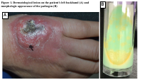
| 抄録 | Buruli ulcer is the third most common mycobacterial infection worldwide and is mainly diagnosed in tropical regions. Globally, this progressive disease is caused by Mycobacterium ulcerans; however, Mycobacterium ulcerans subsp. shinshuense, an Asian variant, has been exclusively identified in Japan. Because of insufficient clinical cases, the clinical features of M. ulcerans subsp. shinshuense–associated Buruli ulcer remain unclear. A 70-year-old Japanese woman presented with erythema on her left backhand. The skin lesion deteriorated without an apparent etiology of inflammation, and she was referred to our hospital 3 months after disease onset. A biopsy specimen was incubated in 2% Ogawa medium at 30 °C. After 66 days, we detected small yellow-pigmented colonies, suggesting scotochromogens. Matrix-assisted laser desorption/ionization time-of-flight mass spectrometry (MALDI Biotyper; Bruker Daltonics, Billerica, MA, USA) indicated that the organism was Mycobacterium pseudoshottsii or Mycobacterium marinum. However, additional PCR testing for the insertion sequence 2404 (IS2404) was positive, suggesting that the pathogen was either M. ulcerans or M. ulcerans subsp. shinshuense. Further examination by 16S rRNA sequencing analysis, focusing on nucleotide positions 492, 1247, 1288, and 1449–1451, we finally identified the organism as M. ulcerans subsp. shinshuense. The patient was successfully treated with 12 weeks of clarithromycin and levofloxacin treatment. Mass spectrometry is the latest microbial diagnostic method; however, it cannot be used to identify M. ulcerans subsp. shinshuense. To accurately detect this enigmatic pathogen and uncover its epidemiology and clinical characteristics in Japan, more accumulation of clinical cases with accurate identification of the causative pathogen is essential.
|
| キーワード | Buruli ulcer
Mycobacterium ulcerans
Mycobacterium ulcerans subsp
shinshuense
16S rRNA sequencing analysis
|
| 備考 | © 2023 Japanese Society of Chemotherapy and The Japanese Association for Infectious Diseases. This manuscript version is made available under the CC-BY-NC-ND 4.0 License. http://creativecommons.org/licenses/by-nc-nd/4.0/.
This is the accepted manuscript version. The formal published version is available at https://doi.org/10.1016/j.jiac.2023.02.009.
This fulltext file will be available in May 2024.
|
| 発行日 | 2023-05
|
| 出版物タイトル |
Journal of Infection and Chemotherapy
|
| 巻 | 29巻
|
| 号 | 5号
|
| 出版者 | Elsevier BV
|
| 開始ページ | 523
|
| 終了ページ | 526
|
| ISSN | 1341-321X
|
| NCID | AA11057978
|
| 資料タイプ |
学術雑誌論文
|
| 言語 |
英語
|
| OAI-PMH Set |
岡山大学
|
| 著作権者 | © 2023 Japanese Society of Chemotherapy and The Japanese Association for Infectious Diseases.
|
| 論文のバージョン | author
|
| PubMed ID | |
| DOI | |
| Web of Science KeyUT | |
| 関連URL | isVersionOf https://doi.org/10.1016/j.jiac.2023.02.009
|
| ライセンス | http://creativecommons.org/licenses/by-nc-nd/4.0/
|