| ID | 69389 |
| FullText URL | |
| Author |
Wang, Xiuting
Department of Periodontology and Endodontology, Tohoku University Graduate School of Dentistry
Suzuki, Shigeki
Department of Operative Dentistry, Okayama University Graduate School, Medicine, Dentistry and Pharmaceutical Sciences
Tsai, Shin-Ho
Department of Operative Dentistry, Okayama University Graduate School, Medicine, Dentistry and Pharmaceutical Sciences
Nagasaki, Karin
Department of Periodontology and Endodontology, Tohoku University Graduate School of Dentistry
Fahreza, Rahmad Rifqi
Department of Periodontology and Endodontology, Tohoku University Graduate School of Dentistry
Omori, Masato
Department of Periodontology and Endodontology, Tohoku University Graduate School of Dentistry
Yamada, Satoru
Department of Periodontology and Endodontology, Tohoku University Graduate School of Dentistry
|
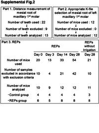
| Abstract | As the pulp regeneration for non-vital teeth is one of the ultimate clinical achievements, regenerative endodontic procedures (REPs) have become the most explored treatment modality. In this technique, periodontal tissue is guided from the apical region into the root canal and pulp chamber to promote attachment. It is well established that immature teeth are effective targets for treatment. However, the indications for this treatment have not yet expanded sufficiently to encompass mature teeth with closed apical apex. In the present study, a mouse model of REPs in mature teeth was established, employing the maxillary first molar mesial root. μCT analyses disclosed that the distance from the occlusal surface to the physiological apex of the maxillary first molar mesial root in mice is 2.14 mm ± 0.08 mm, and the distance from the occlusal surface to the periapical alveolar bone is 2.46 mm ± 0.10 mm. Mesial root canal was treated with several sizes of k-files, and 15# k-file was identified as the most suitable k-file for use (P = 0.0007). During the regenerative process, spindle-shaped fibroblast-like cells, fibrous tissue formation, and mineralized tissue formation were identified on days 14 and 28. This study demonstrated that it is feasible to use the maxillary first molar mesial root as a REPs model for mature teeth and provided a detailed protocol and analysis of the healing process.
|
| Keywords | Regenerative endodontic procedures
Establishment of protocols
Mouse experimental model
Mature teeth
|
| Published Date | 2025-09-29
|
| Publication Title |
Odontology
|
| Publisher | Springer Science and Business Media LLC
|
| ISSN | 1618-1247
|
| NCID | AA11580928
|
| Content Type |
Journal Article
|
| language |
English
|
| OAI-PMH Set |
岡山大学
|
| Copyright Holders | © The Author(s) 2025
|
| File Version | publisher
|
| PubMed ID | |
| DOI | |
| Related Url | isVersionOf https://doi.org/10.1007/s10266-025-01211-4
|
| License | http://creativecommons.org/licenses/by/4.0/
|
| Citation | Wang, X., Suzuki, S., Tsai, SH. et al. Establishment of a regenerative endodontic procedures model of mature mouse teeth and evaluation of the wound healing process. Odontology (2025). https://doi.org/10.1007/s10266-025-01211-4
|
| 助成情報 |
( Okayama University )
|